Christine U. Lee1, Jin Ye Yeo2
1Mayo Clinic, Department of Radiology, Division of Breast Imaging and Intervention, Rochester, MN 55905, USA; 2TBCR Editorial Office, AME Publishing Company
Correspondence to: Jin Ye Yeo. TBCR Editorial Office, AME Publishing Company. Email: tbcr@amegroups.com
This interview can be cited as: Lee CU, Yeo JY. Meeting the Editorial Board Member of TBCR: Dr. Christine U. Lee. Transl Breast Cancer Res. 2024. https://tbcr.amegroups.org/post/view/meeting-the-editorial-board-member-of-tbcr-dr-christine-u-lee.
Expert introduction
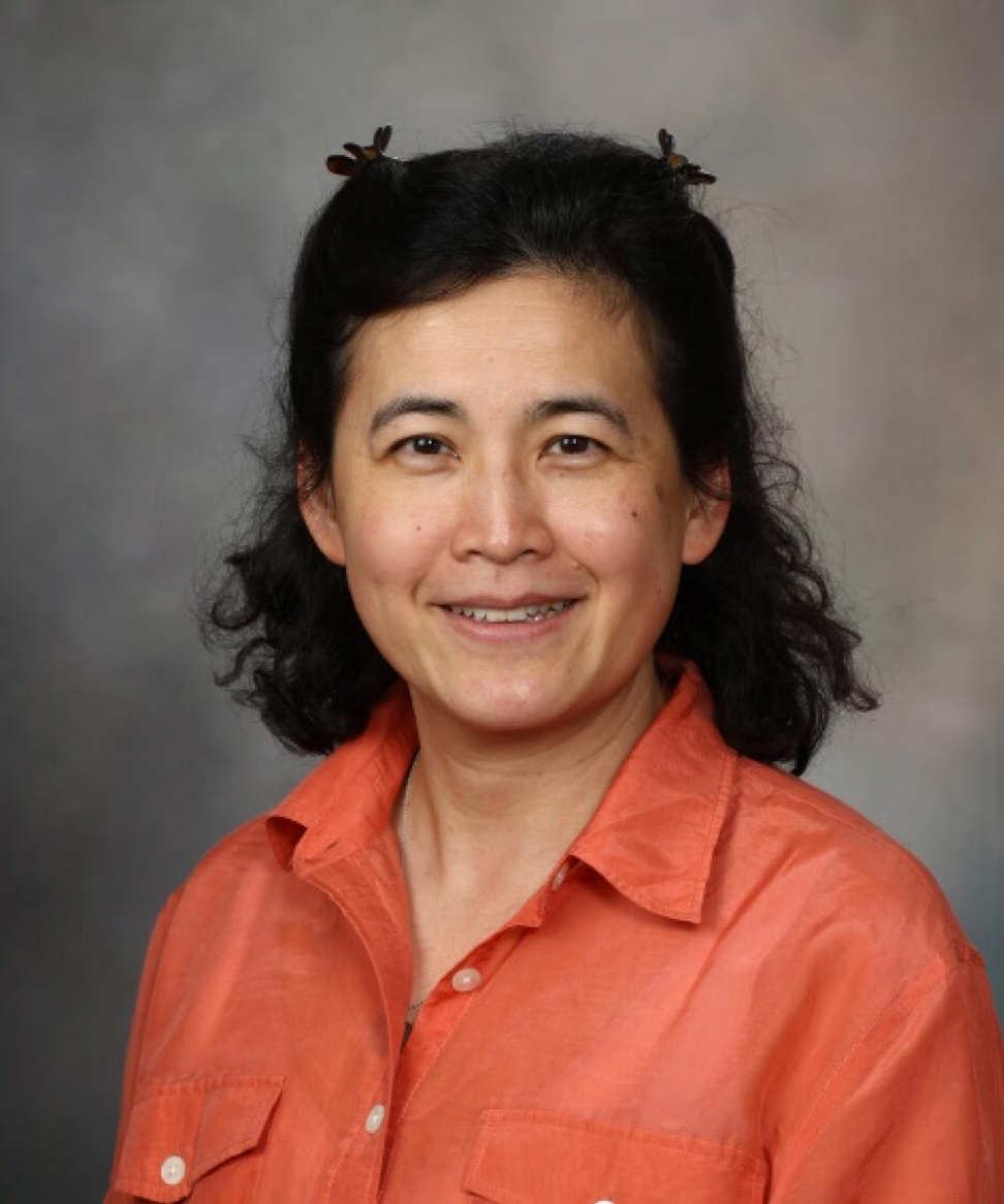
Dr. Christine U. Lee (Figure 1) earned her M.D.-Ph.D. degrees and completed her radiology residency and cross-sectional fellowship training at the Mayo Clinic in Rochester, MN, USA. She is a board-certified radiologist with nearly two decades of post-training experience in breast radiology. She has played a pivotal role in medical education, training and mentoring numerous residents and fellows. She has been recognized with the Teacher of the Year Award twice for her exceptional teaching contributions. Her mentees have received research awards and funding including the Radiological Society of North America (RSNA) Resident Research grant. Dr. Lee has co-authored an 881-page book on body MRI.
Dr. Lee is an NIH-funded researcher engaged in developing an ultrasound-detectable, migration-resistant biopsy marker for improving care in patients with breast cancer. Her contributions extend to other advancements and innovations, including contrast-enhanced ultrasound lymphography, which improves the identification of superficial lymphatic channels in patients with extremity lymphedema anticipating lymphovenous bypass surgery. Additionally, she has established a framework and guide for administering intralesional steroid injections in patients with idiopathic granulomatous mastitis.

Figure 1 Dr. Christine U. Lee
Interview
TBCR: What initially inspired you to pursue a career in breast radiology?
Dr. Lee: Early in my career, I worked as a cross-sectional radiologist interpreting body magnetic resonance imaging (MRIs), body ultrasounds, and abdominal computed tomography (CTs), and performing procedures. I can remember the days when breast MRIs were still performed and interpreted by body radiologists. At one point, our breast radiology division became short-staffed, and I was asked to help out and I never left. I discovered that breast radiology offered opportunities and a special blend of the imaging modalities I enjoyed, such as x-ray (mammograms), MRIs, ultrasound, and procedures. I also found the collaborative nature and camaraderie of working with multidisciplinary teams in patient care particularly rewarding.
TBCR: Could you provide a brief overview of the advancements in breast imaging radiology? What are some examples that have made a significant impact?
Dr. Lee: During my career, several notable advancements in breast imaging have made a significant impact. One major shift was the transition of breast MRI from the body MRI practice to the breast radiology practice. We have also been a part of the transition from film-screen mammography to digital mammography, and now to digital breast tomosynthesis. Our role in de-escalating axillary surgery in the setting of node-positive breast cancer continues to evolve. Other growing advancements in the field include contrast-enhanced mammography, artificial intelligence, and its effective and ethical integration into breast imaging practices.
TBCR: Your NIH-funded research focuses on developing an ultrasound-detectable, migration-resistant biopsy marker. Can you explain how this innovation could improve patient care in breast cancer?
Dr. Lee: For many of us breast radiologists, we are thrilled when patients with breast cancer and node-positive disease or axillary metastasis respond favorably to neoadjuvant therapy. However, it can be challenging to preoperatively localize the treated axillary lymph node, which often appears normal after treatment and has a biopsy marker that is hard to detect by ultrasound. Our biopsy marker leverages a nearly three-decade-old imaging feature called color Doppler twinkling, commonly associated with kidney or urinary stones, to identify it. Preliminary results from our clinical trials are promising, with some surgeons now able to use ultrasound to locate the marker themselves, sparing patients an additional invasive localization step and reducing costs. Surgeons can also use twinkling to confirm that the marker has been resected, reducing operating room (OR) and intubation time, costs, and patient morbidity.
TBCR: Could you elaborate on your work with contrast-enhanced ultrasound (CEUS) lymphography and its impact on managing extremity lymphedema?
Dr. Lee: Over the years, we have collaborated closely with our plastic surgeons to develop CEUS lymphography into a clinical exam. CEUS lymphography can identify potential superficial lymphatic vessels suitable for lymphaticovenous anastomosis (LVA) surgery. While indocyanine green (ICG) lymphography with near-infrared (NIR) fluorescence imaging is limited to superficial depths, CEUS lymphography can identify lymphatic candidates more than a centimeter deep to the skin, even when a lymphatic stardust pattern is present. CEUS lymphography offers a chance to identify more candidates for LVA surgery. There is still room for improvement to better serve this patient population.
TBCR: You are also working on establishing a framework for intralesional steroid injections in idiopathic granulomatous mastitis (IGM). What challenges have you faced in this area, and what do you hope to achieve with this framework?
Dr. Lee: I still remember feeling defeated by my first case of advanced IGM back in 2015. At the time, we were not even certain it was IGM, but we have learned a lot since then. Patients with IGM often endure debilitating pain and develop skin fistulas, which can be intractable despite antimicrobial therapy and systemic steroids. While intralesional steroid injections had been described, there was not a clear framework for characterizing the extent of disease and how to dose the steroids accordingly. The framework we developed aims to provide more structure for both evaluating the severity of IGM and administering intralesional steroid injections in a more systematic way.
TBCR: As an educator recognized for your exceptional teaching contributions, how do you approach mentoring your trainees to help them achieve success in their research and clinical careers?
Dr. Lee: I always tell trainees that it is more important for them to understand why they are doing something rather than just mimicking my approach. They do not have to do things exactly the way I do, but I encourage them to keep it in mind as an option to consider when they run into a complex case in the future. For example, I once had a resident whom I was teaching how to do a fine-needle aspiration with a syringe. While I could hold the syringe in a way to actively aspirate, the resident had much larger hands than mine. But, because he understood the reasoning and the ultimate goal, he was able to modify the technique to make it work for him.
TBCR: How has your experience been as an Editorial Board Member of TBCR?
Dr. Lee: As an Editorial Board Member, I have been delighted to be a part of its growth. The TBCRteam is extremely responsive, and the scope of TBCR effectively encompasses the multidisciplinary specialties involved in breast cancer and breast disease. Because of this, it has been a pleasure to recommend TBCR to colleagues for manuscript submission, and they consistently find the journal to be a good fit. Overall, being an Editorial Board Member has been a rewarding experience.
TBCR: As an Editorial Board Member, what are your expectations for TBCR?
Dr. Lee: I would like to see TBCR continue to grow to be a formidable journal in this field. By incorporating multidisciplinary perspectives, the journal fosters a more comprehensive approach to advancing and translating breast cancer research and care.
